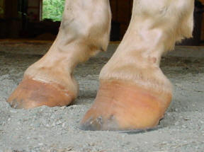
Club Feet Prevention, Treatment & Correction Anyone who has spent any time with equines has undoubtedly seen club feet A club foot horse is typically recognized and defined as having one front hoof growing at a much steeper angle than the other, with a short dished toe, very high heels, extremely curved wall and straight bars The club foot is also generally much When the foot is unbalanced, and then shod, the horse tends to compensate by twisting the foot This eventually will put undue stress on the hock from the torque of twisting the joint And by unbalanced I would guess, in this case, that the inside of the foot (the wall) is higher than the outside which is typical for hind feet Navicular syndrome is a common condition, but it is not simple or straightforward To better understand why it seems like one treatment works great for one horse and marginally for another, it is important to understand a little bit of the history of navicular syndrome, the anatomy that is involved, and available treatment options
Boarding At Yucca Veterinary Medical Center
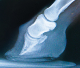
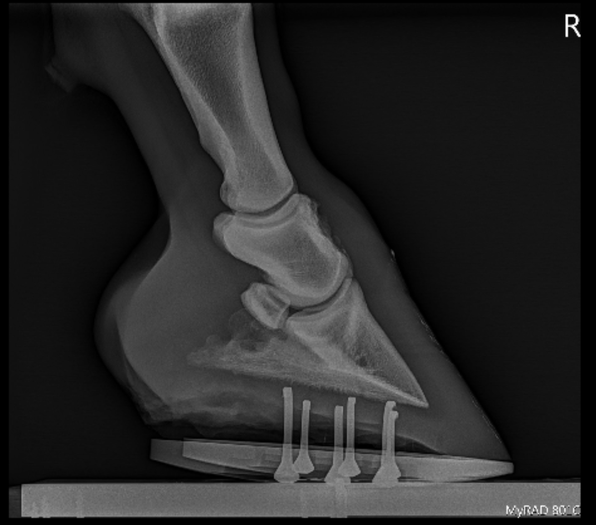
Club foot horse radiograph
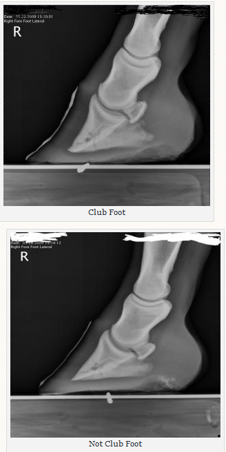
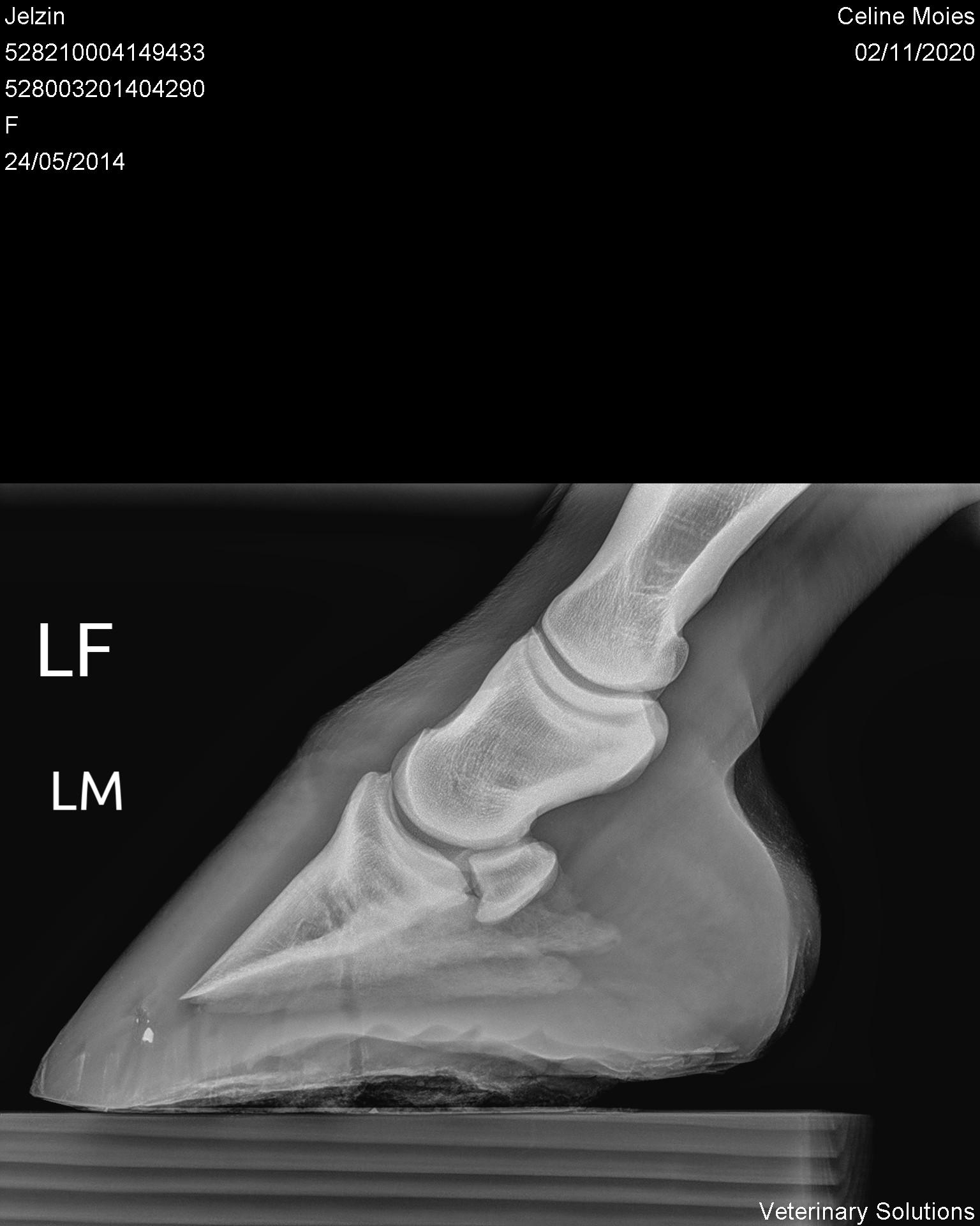
Club foot horse radiograph- club foot case recently This particular horse, a six year old gelding, has what I feel is a grade three club foot (on a 15 scale) Apparently the club foot condition has been with this horse since it was a foal This horse found it difficult toClub foot refers to a hoof that is more upright than normal It is often associated with a concave front (dorsal) hoof wall, high (often contracted) heels, and widening of the white line from mechanical stretching of the hoof wall attachments (the laminae) Adult club foot requires a completely different approach to treatment than juvenile club foot


Clubfoot Barefoot Hoofcare
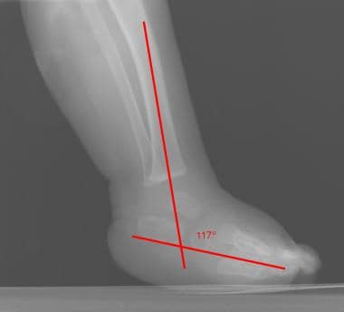
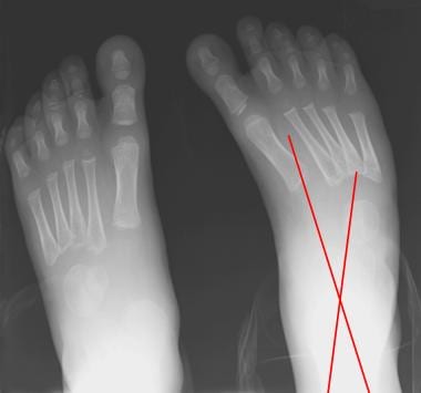
Reversing Distal Descent of P3 () Pete Ramey In the healthiest of equine feet, the hoof walls should be firmly attached to the coffin bones and the coronet should lie at the same level or even slightly below the coffin bone (P3) This allows proper uninhibited motion of the coffin joint It also ensures the horse can have a naturally Radiographs show the true gain in foot (ankle) dorsiflexion and confirm the appearance of an iatrogenic rockerbottom foot should one result Occasionally, radiographs are necessary to diagnose clubfeet associated with tibial hemimelias Talocalcaneal parallelism is the radiographic feature of clubfoot (talipes) Club Foot Horses Versus Uneven Weight Distribution First off let's discuss exactly what a "club foot" is This term is widely misused with regard to its use in horses with uneven hoof growth patterns The term "club foot" actually refers to a congenital defect of the foot and according to The Free Dictionary,
In older horses, when club foot has become a more chronic condition, altering the biomechanics of the foot is often the key to management Radiographs of the feet are essential in providing the information necessary to evaluate the existing biomechanics and determining the appropriate changes that need to be madeAny club foot that has been around a while will have a sensitive, unused, underdeveloped frog/digital cushion You can fix everything else and still have the back of the foot too sensitive for the horse to land on, which will cause the shortened stride and resulting club foot on its own – another vicious cycleOf club foot A horse with club foot has one hoof that grows more upright than the other The "up" foot is accompanied by a broken forward pastern, that is, the hoof is steeper than the pastern (Photo 1) In a normal foot, the hoof capsule and the pastern align
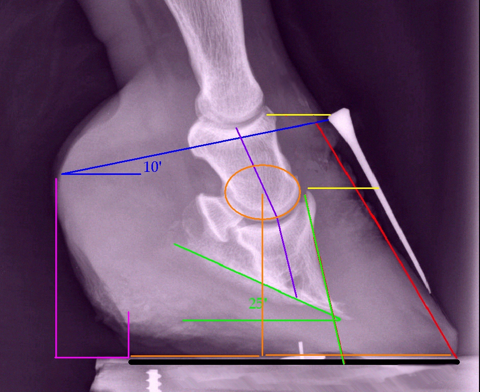
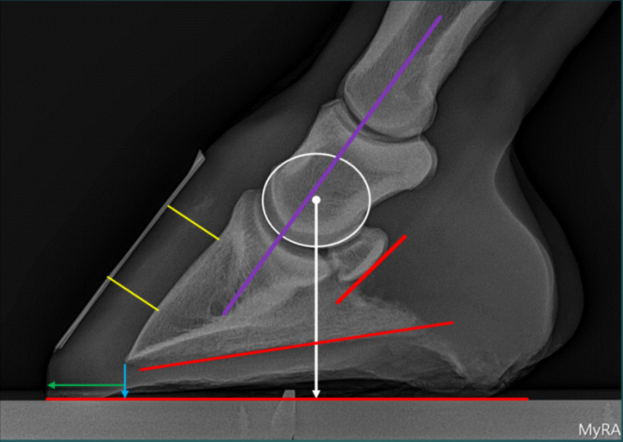
Labels equine, farrier, hoof, hoof specialist, how to take radiographs, normal horse hoof radiograph, Podiatry, radiographs, Sammy L Piittman Monday, Whats in a toe angle Whats in a toe angle?Large hoof wall to distal phalanx distances (HDPD) have been observed in horses with chronic laminitis in the absence of other radiological indicators such as distal phalanx rotation or sinking 1 However, the upper limit of 'normal' is debatable, and so the aim of this retrospective study was to document the HDPDpalmar length of the distal phalanx (HDPD ratio) in a range of horses and There are few studies on the correlations between radiographic measurements of the foot and abnormalities of specific structures found with magnetic resonance imaging (MRI) Objectives To document the relationship between radiographic measurements of the equine foot and the presence of lesions in the foot on MRI


2


2
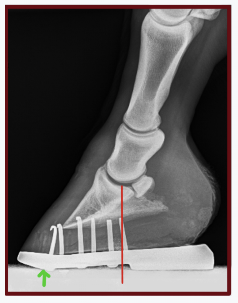
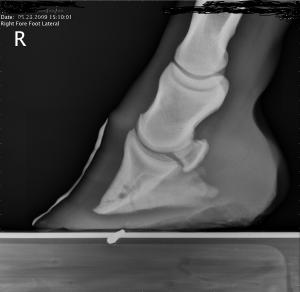
A club foot alters a horse's hoof biomechanics, frequently leading to secondary lamenesses Affected horses tend to land toefirst, and their heel's growth rate is• rotatory subluxation of talocalnoenavicular joint (subtalar) complex with talus in plantar flexion and subtalar complex in medial rotation and inversion • also reffered as clubfoot • talipes derived from term talus ankle & pes foot • equinovarus derived from word equino like a horse & varus turned inward 4Comes very useful in horses with upright feet, the best example being the clubfooted horse As the foot grows out in these horses, there is a propensity for the dorsal wall to distort and flare, producing multiple angles to the dorsal wall Radiographic evaluation of the dorsal wall with a conforming marker allows accurate assessment of the distortion
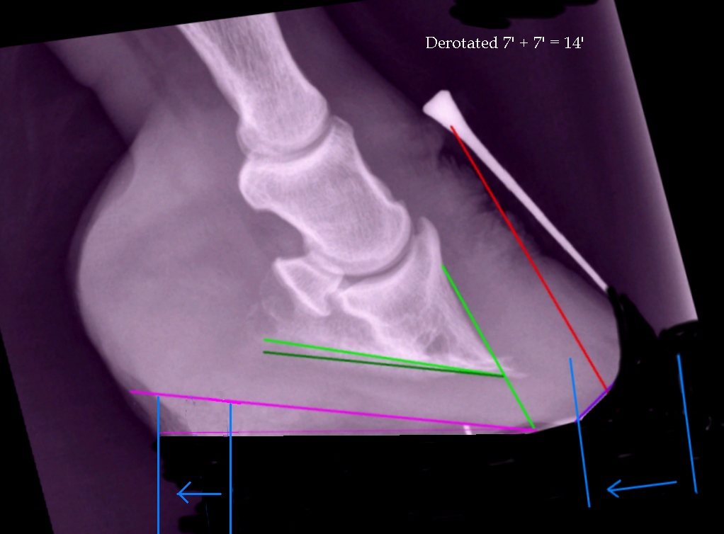


High Heels The Laminitis Site


Hoof Evaluation Radiographs For The Farrier
The equine club foot is defined as a hoof angle greater than 60 degrees What we see externally as the equine clubbed foot is actually caused by a flexural deformity of the distal interphalangeal joint (coffin joint) Causes include nutritional issues, heredity, position in the uterus or injury The condition is most often encountered in young animals and can be either congenital (they are bornUsually, club foot affects both front legs with one being more severe than the other Open all Close all Plus sign () if content is closed, 'X' if content is open The foot with less weight on the heel tends to develop a longer heel A mild club foot may worsen if trimmed infrequently or improperly and by the time the horse is 2 or 3 years old the problem becomes more obvious The horse may develop an uneven stride and rough gait (and some loss of agility) due to the mismatched hoof angles


Hoof Conformation Vs Horse Conformation Scoot Boots Retail


The Progressive Equine Services Hoof Care Centre Facebook
The lateral radiograph confirmed how little foot we really had to work with, which the horse's pain level and hoof inflammation was certainly telling us What was also very interesting was the DP radiograph, since it also indicated significant medial lateral hoof imbalances that we could help this horse with at the time A horse that tends habitually to put the same foot out front all the time needs a closer diagnostic look He referred to a study in the Netherlands ofThat's a completely different foot inside even though it has the same hoof angle


Shoeing Options For Club Foot In Horses


Equine Foot Radiographs Views Navicular
My horse has a club foot Can it be treated?It is hoped that MRI will validate certain radiographic changes seen on the standard foot examination Radiographic examination of the digit will remain the common imaging technique between the field practitioner and the referral hospital inWhen doubt persists, the horse should be confined and a second radiographic examination carried out 14 to 21 days later After this interval a fracture appears wider and more irregular Increased vascular channels may be seen in Thoroughbreds without generating lameness or local pain
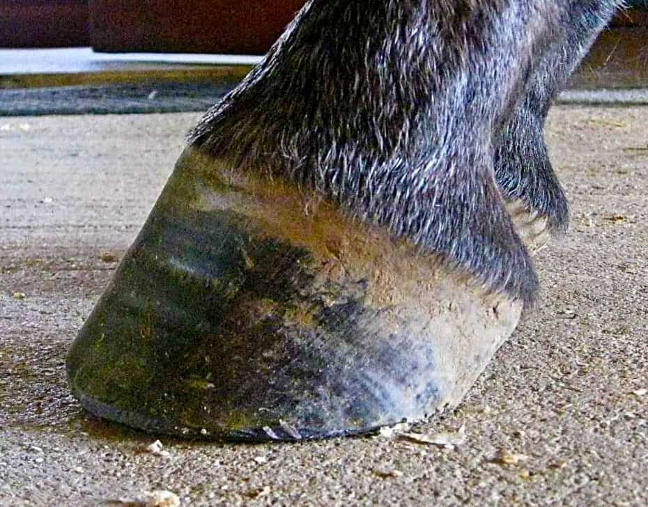


The Tolerable Club Foot The Horse


What S New In Equine Foot Radiology Imv Imaging
This horse came up acutely "fracture lame" with swelling in this area of the leg, which caused the vet to question whether this line was really normal, or if the horse had been injured In the end, the horse had a softtissue injury completely unrelated toSurgery often a necessity for a horse with a club foot Flickrcom Smerikal There are three general causes of club feet genetic, nutritional, and grazing stance (with one foot forward and one back) – and a combination of these Club feet are more common in some breeds and in specific bloodlinesRadiographic Examination of the Equine Foot W Rich Redding Radiography and radiology of the equine foot R Weller 50th BEVA Congress 11 O'Brien's Radiology for the Ambulatory Equine Practitioner Timothy R O'Brien (05) Atlas of Diagnostic Radiology of the Horse Diseases of the Front and Hind Limbs Kees J Dick and Ilona Gunsser


Lesson 4


The Wild Mustang Hoof
CLUB FOOT 8 CLUB FOOT Definitions Talipes Talus = ankle Pes = foot Equinus (Latin = horse) Foot that is in a position of planter flexion at the ankle, looks like that of the horse Calcaneus Full dorsiflexion at the ankle 9Here are two foals that would be considered to have a club foot with a toe angle of close to 64 degrees However what makes up theXray of feet (typical clubfoot) Clubfoot Introduction Clubfoot (talipes equinovarus – TEV) is one of the major orthopedic conditions of childhood One of the most common of all birth defects, clubfoot affects about 1 in 400 babies born in the United States each year Boys are affected twice as often as girls Causes


Boarding At Yucca Veterinary Medical Center


3
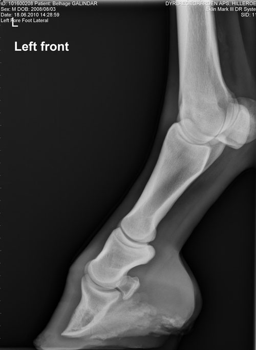
Diagnosis of Club Foot in Horses Observation is the most efficient method of diagnosis whereby the constant checking of your newborn foal’s hooves would be advisable Birth deformities are usually easy to notice, especially if your foal has difficulty standing to nurseAny foot radiograph for alignment First, you can evaluate the relationship of the tibia to the hindfoot, then the relationship of the hindfoot to the midfoot, and finally the relationship of the midfoot to the forefoot As you read the case scenarios, you will see how this can be aMajor sources of coffin joint pain include a broken forward hoof axis (mild club foot) and mediallateral hoof imbalance Wedge tests Frog wedge To do this test, a board or hoof knife is placed under the frog and the horse made to stand on it for 1 minute by lifting the other foot


Clubfoot Barefoot Hoofcare


How To Treat Club Feet And Closely Related Deep Flexor Contraction
The name of the view describes the direction of the xray beam The beam is aimed from dorsoproximal to palmarodistal at a 65 degree angle to the sole of the foot This view may be obtained with the horse standing on the cassette as in this illustration The xray beam is centered at the coronary band Notice in the photo that the cassetteFetlock is a term used for the joint where the cannon bone, the proximal sesamoid bones, and the first phalanx (long pastern bone) meet The pastern is the area between the hoof and the fetlock joint Disorders of the fetlock and pastern include conditions such as fractures, osteoarthritis, osselets, ringbone, sesamoiditis, synovitis, andAnd variable angle horizontal beam dorsopalmar foot radiographs were acquired while keeping the limb position constant Analyses of measurements demonstrated that hoof pastern angle had a linear relationship (R 2 = 0, P < 0


Farrier Terms Decoded Top 5 Farriery Terms Horse Owners Should Know Badger Veterinary Hospital


Hoof Deformities Round Pen Square Horse
Dorsopalmar foot radiographs were acquired while varying the lateromedial stance; Clubfoot, or talipes equinovarus, is a congenital deformity consisting of hindfoot equinus, hindfoot varus, and forefoot varusThe deformity was described as early as the time of Hippocrates The term talipes is derived from a contraction of the Latin words for ankle, talus, and foot, pesThe term refers to the gait of severely affected patients, who walked on their ankles Radiographic examination of the foot is one of the most frequently performed studies of equine limbs 2 It is somewhat more complex than other radiographic studies of horse limbs, but with proper equipment and technique excellent quality radiographs can be made Without a good quality radiographic examination of the foot, correct radiographic diagnoses cannot be
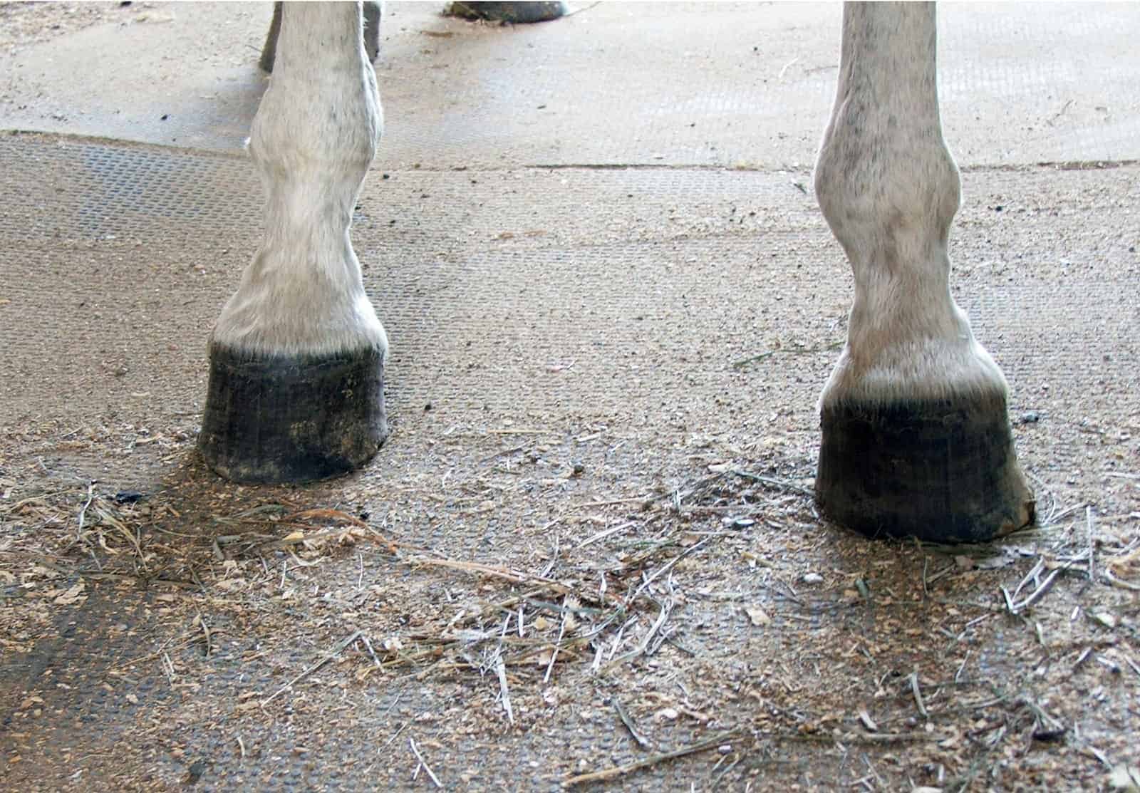


Club Foot Heritability In Horses The Horse


2
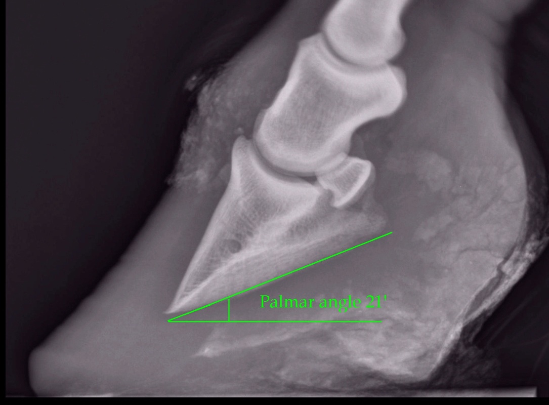
The xray will show whether the hoof pastern axis is parallel If the axis is broken forward (club foot) or if the axis is broken back (long toe underrun heel), the radiograph will reveal the degree of deformity and the best way to trim the foot to improve it Using landmarks, measurements can be drawn on the radiographs and transferred to the footThe second type of situation I see is a good foot that holds a positive palmar angle, good sole depth and typically has a stronger bone angle This group will often be bilateral lame or more lame in the club (higher profile heel) foot, have a shortened, choppy, stabbing gait and blocks to a low palmar digital nerve block Club Foot Conformation in Horses Caused by abnormal contraction of the deep digital flexor tendon, a club foot puts pressure on the coffin joint and initiates a change in a hoof's biomechanics Telltale signs of a club foot may include an excessively steep hoof angle, a distended coronary band, growth rings that are wider at the heels


Clubfoot Barefoot Hoofcare


Equine Podiatry Say What Mobile Veterinary Services
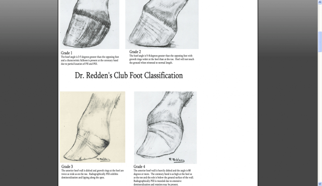
A horse with an upright alignment of the pastern bones will also have upright hoovesa situation that is sometimes mistaken for club foot A true club foot is significantly more upright than the other hooves, or the angles of both hoof walls are steeper than the angles of the pasterns The severity of the problem is commonly graded on a fourpoint scale Grade 1, theAnce of the foot should be determined and the foot should be inspected for underrun or separated wall or sole Sensitivity to hoof testers or response to firm pressure from fingers should be assessed If lameness is present, peripheral nerve blocks should be performed to isolate and confirm the origin of the lameness Radiographs of the foot shouldTwo studies evalu ated the effect of horse stance on measurements of the hoof capsule or phalanges 45 In the first, lateromedial radiographs of the front feet of five horses were acquired with the metacarpal region positioned vertically, and at various degree s angled cranially and caudally, as well as with the forelimbs only positioned on raised blocks 4 The angle of the solear


Recognizing And Managing The Club Foot In Horses Horse Journals


Equine Podiatry In Wendell Nc Neuse River Equine Hospital
The horse's feet need to be picked out and wire brushed clean, including the hoof wall from ground surface to the coronary band, around the heels, into the collateral groves, central sulcus, and any other separations and pockets, for clear visibility of all structures in the radiographBoth hooves have a 62° hoof angle The first foal's club foot has a slight dish in the front, a high PA of 12°, and a BA of 50° That foot might be a candidate for check ligament desmotomy But the other foal's club foot has a 23° PA and a BA of 60°; A horse with a club foot is kind of like a horse in high heels The hoof angle becomes raised and the horse walks on his toe due to a shortening of the musculotendinous unit (the unit including


Surgical Services Scenic Rim Veterinary Service


Michael Porter Equine Veterinarian 12


Understanding Club Foot The Horse Owner S Resource
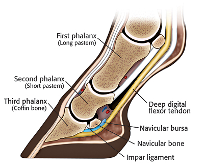


The Chronicle Of The Horse


2


2


Hoof Evaluation Radiographs For The Farrier


Natural Angle Volume 15 Issue 1 Spanish Lake Blacksmith


Zangersheide Breeders Sales Tweety Van De Lentakker A T Z
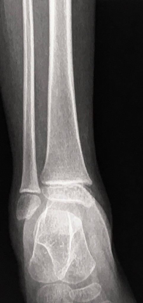


Equinus Foot Deformity Radiology Case Radiopaedia Org


Recognizing Various Grades Of The Club Foot Syndrome


1


Understanding X Rays The Laminitis Site


Shoeing Options For Club Foot In Horses


2


Radiographic And Radiological Assessment Of Laminitis Sherlock 13 Equine Veterinary Education Wiley Online Library
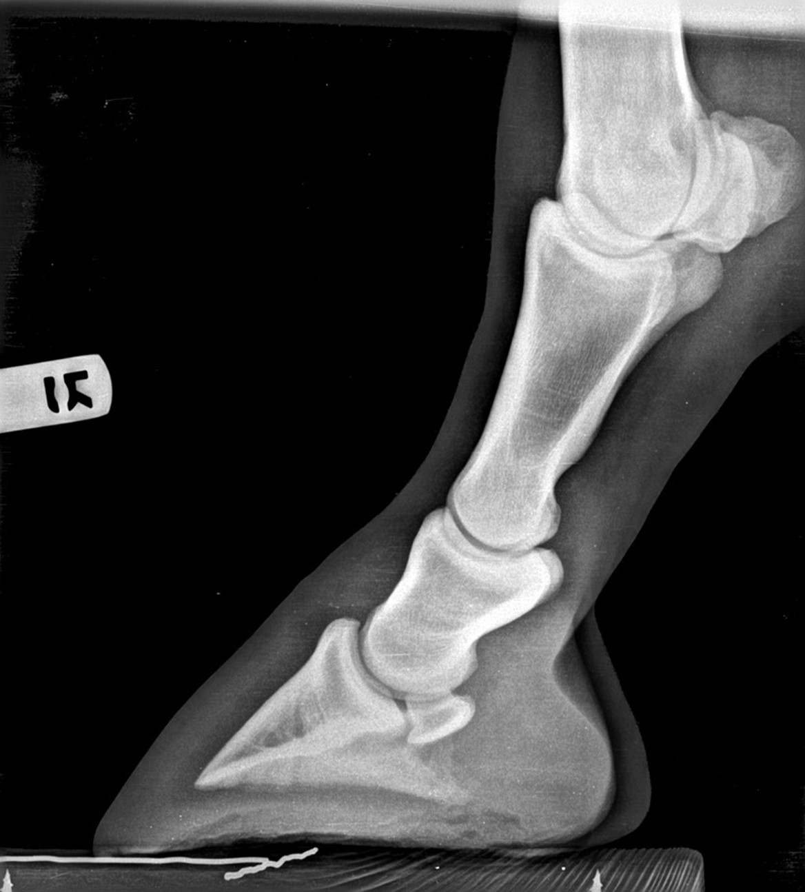


Hoof Radiographs Springhill Equine Veterinary Clinic


Club Foot In Horses Brian S Burks Fox Run Equine Center Facebook


Belgian Horse Trading 5 Jaguar Ms


No Foot No Horse Part 2 Hayes Equine Veterinary Services


Recognizing Various Grades Of The Club Foot Syndrome


2


2


Radiolucency Glue On Horseshoes By Sound Horse Technologies


Laminitis A Pictorial Review


Equine


Farriery For The Hoof With A High Heel Or Club Foot Semantic Scholar


Equine Therapeutic Farriery Dr Stephen O Grady Veterinarians Farriers Books Articles


A Lateral And B Dorsopalmar Venograms Of A Normal Foot Areas Of Download Scientific Diagram


Foal With Hoof Problems Club Foot The Horse
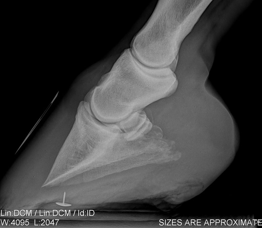


Understanding X Rays The Laminitis Site


Imaging Anatomy


Equine Podiatry Dr Stephen O Grady Veterinarians Farriers Books Articles


Thrush Cured Cowboy Pads And Copper Sulfate
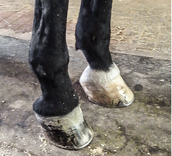


What Advice Has Been Most Helpful When You First Encounter A Club Foot American Farriers Journal


Managing The Club Hoof Easycare Hoof Boot News


1


The Wild Mustang Hoof


Recognizing And Managing The Club Foot In Horses Horse Journals


Problem Cases Steel Shoes Farrier Service


How To Manage The Club Foot Birth To Maturity The Horse


High Heels The Laminitis Site


Equine Therapeutic Farriery Dr Stephen O Grady Veterinarians Farriers Books Articles


Club Foot Or Upright Foot It S All About The Angles American Farriers Journal


Recognizing And Managing The Club Foot In Horses Horse Journals


Understanding Navicular Syndrome Heel Pain In Horses


Club Foot


Cur Ost Normal And Not So Normal Horse Feet


The Progressive Equine Services Hoof Care Centre Facebook


Clubfoot Imaging Practice Essentials Radiography Computed Tomography


Clubfoot Barefoot Hoofcare


Managing The Club Hoof Easycare Hoof Boot News


Belgian Horse Trading 11 Jelzin


Foal With A Club Foot Download Scientific Diagram


2


Severe Lameness Scarsdale Vets


Club Foot Horses Club Foot Feet Club


Low Foot Case Study Dixie S Farrier Service


Laminitis Signalment Treatment And Prevention Flying Changes


Clubfoot Barefoot Hoofcare


New Strategies For Treating Club Foot From Foal To Adult


Epublishing


Podiatry Burwash Equine Services


Hoof Angles Part 5 Enlightened Equine


Hoof Experts X Rays Included The Horse Forum


2


Recognizing And Managing The Club Foot In Horses Horse Journals


Understanding X Rays The Laminitis Site


Contact Us Palmetto Equine Veterinary Services


Basic Shoeing Working With A Club Foot Farrier Product Distribution Blog


Podiatry Burwash Equine Services


Hoof Evaluation Radiographs For The Farrier


Recognizing And Managing The Club Foot In Horses Horse Journals


Clubfoot Imaging Practice Essentials Radiography Computed Tomography


Zangersheide Breeders Sales Stella De Betze Z


2


Comstock Equine Hospital Veterinarian In City State Country Corrective Trimming


2


Michael Porter Equine Veterinarian 12


Farriers Archives Springhill Equine Veterinary Clinic


0 件のコメント:
コメントを投稿